Magnetic Resonance Imaging (MRI): An MRI does NOT use X-rays or other ionizing radiation to image patients.
Ultrasound (US): An Ultrasound does NOT use X-rays or other ionizing radiation to image patients.
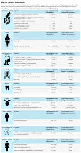
X-ray: A small dose of radiation is used to perform most X-ray examinations. As an example, the radiation exposure from a typical chest X-ray is comparable to the radiation exposure received during a cross-country plane trip.
Mammogram: A small dose of radiation is used to perform this study – approximately equal to the radioactivity your own body naturally produces each year.
Computed Tomography (CT): A CT scan requires more radiation than conventional X-ray examinations; however, it also provides more detailed pictures. Total radiation exposure varies greatly by CT procedure; a typical chest CT is comparable to the radiation exposure the average American receives from radon gas naturally occurring in their home.
Positron Emission Tomography (PET): Positron Emission Tomography uses small amounts of radioactive isotope-labeled materials which are injected to target the area of the body being imaged. The radiation dose varies by procedure.
Please visit our Imaging Questions and Precautions page for more information on things to discuss with your doctor, radiologist or technician.
According to recent estimates, the average person in the U.S. receives an effective dose of about 3mSv per year from naturally occurring radioactive materials and cosmic radiation from outer space. These natural “background” doses vary throughout the country.

