


Women’s health experts recommend that women with no breast symptoms or cancer risk factors start screening mammograms at the age of 40 and repeat them annually (every 12 months). For women with high risk factors such as family history, your physician may recommend starting screenings sooner.
• Expect to spend 45 minutes to an hour at the facility to complete the entire process.
• Please do not wear deodorant, lotion, powder, or perfume under your arms or on your breasts the day of the procedure.
• Prior to your exam you will be asked to undress from the waist up. A gown will be provided for your comfort. It is best if you wear shorts, pants, or a skirt to your appointment so that you can easily undress from the waist up.
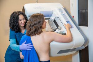
• The mammography technologist will interview you and review your medical history.
• The technologist will visually inspect your breasts for any skin changes or puckering. They may also place markers on your skin showing where moles and/or surgical scars are located.
• The technologist will manually place your breast in the machine for each image to ensure proper positioning while also coaching you for body placement to ensure you are as comfortable as possible.
• There are 4 standard images that will be obtained: 2 of each breast. Additional images may be necessary in the event that the image quality was not optimal, if you have breast implants, or if the entire breast cannot be seen on a single image.
• For each image the breast will be compressed between two plates in order to obtain the best possible image.
• Breast compression is necessary to obtain the best possible images while exposing patients to the smallest possible amount of radiation.
There is no evidence to show that the current level of radiation exposure for annual mammograms will increase the risk of breast cancer. This is considered a low dose and well below the FDA established threshold of acceptable radiation dose. Radiology Ltd., as well as all imaging centers performing mammograms, are held to specific guidelines with frequent equipment inspections to ensure that the equipment is safe and uses the lowest radiation dose possible to produce high quality images. Bottom line, the benefits of early breast cancer detection and treatment far outweigh any theoretical harm from the low dose radiation exposure.
Learn how 3D Mammography Works from our Demonstration Video
It’s estimated that nearly 50% of all women have dense breast tissue. Dense breast tissue is a term used to describe how breast tissue appears on a mammogram. On the images, non-dense (fatty) tissue appears darker, while dense tissue appears white. Because dense tissue shows up as solid white areas, it can sometimes make mammogram images more difficult to interpret. These areas may overlap normal breast structures and, at times, make findings harder to see.
While mammography remains one of the most effective tools for early breast cancer detection, your radiologist may recommend additional imaging (such as ultrasound or MRI) if you have dense breast tissue. These supplemental exams can provide clearer views and help ensure the most complete evaluation possible.
After your study, the images will be evaluated by one of our board-certified radiologists with expertise in breast imaging. A final report will be sent to your doctor or healthcare provider, who can then discuss the results with you in detail.
Reports are also available on the Patient Portal
Receiving an abnormal screening mammogram result can understandably cause worry, but it’s important to know that an abnormal result does not necessarily mean breast cancer.
An abnormal or unclear finding simply means there is an area of the breast that looks different from what is typically seen, or from your prior mammograms. In these cases, additional imaging is recommended to take a closer look and better understand what’s being seen. This may include breast ultrasound, breast MRI, or, in some situations, a breast biopsy.