Skip to Content
Procedures

CT Colonography

What is CT Colonography (Virtual Colonoscopy)?

CT colonography uses X-ray pictures taken from multiple different angles around the body to create cross-sectional images that appear as “slices” of the organs in the abdomen and pelvis. When the colon is distended with gas, these images can be viewed on a special computer workstation that allows the inner walls of the colon to be evaluated in a manner similar to colonoscopy, which is a more invasive procedure.

CT colonography is a safe screening tool that can be used to detect colon polyps in individuals with a relatively low risk of colon cancer. CT colonography is much less invasive than colonoscopy; however, if a large (1 cm or larger) polyp is found, a regular colonoscopy examination may be recommended in order to allow the polyp to be removed. If a smaller (between 6-9 mm) polyp is found, a follow up CT colonography examination in 3 years may be recommended to follow up on the size of the smaller polyp, as these smaller polyps have a less than 1 percent chance of becoming cancerous. You should discuss the pros and cons of CT colonography with your gastroenterologist or other healthcare provider to determine which screening procedure is most appropriate for you. You should also be aware that CT colonography is still considered by some to be an investigational study and may not be covered by your insurance carrier.

You are not a candidate for CT colonography if:

• You have acute diverticulitis (the procedure should not be performed until you have had 4-6 weeks of treatment and your symptoms have resolved).

• You have active inflammatory bowel disease (ulcerative colitis or Crohn’s disease).

• You have a colostomy bag.

How Should I Prepare?

At least 48 hours prior to your procedure, you will need to pick up a prep kit from Camp Lowell Imaging Center (our only center where CT colonography is performed). A technologist will go over all of the instructions and will answer any questions you may have. The prep starts with a low-residue diet two days before the exam, with a liquid-only diet and a bowel prep consisting of stool softeners, laxatives, and barium contrast agents the day before your exam.

You should continue medications prescribed by your doctor or healthcare provider unless informed otherwise. Diabetic patients may need to delay their medication on the morning of the exam until after they have eaten in order to avoid an insulin reaction.

What Should I Expect?

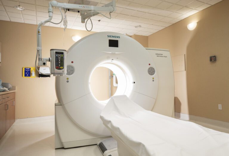
After you arrive for your appointment, you will be escorted to a procedure room, where you will be asked to change into a patient gown. You should remove all jewelry and other removable items such as glasses, dentures, and hearing aids. Women should always inform their technologist if there is any possibility of pregnancy. You will not be sedated for this exam because you will need to follow breathing instructions while the images are obtained.

During the exam you will lie on a table that moves into the doughnut-shaped scanner. A small tube will be inserted into the rectum, and a balloon will be inflated to help hold the tip in. The colon will then be filled with carbon dioxide gas, which can cause feelings of abdominal fullness, cramping, and discomfort that can last for several hours after the exam is completed. This inflation is necessary so that the radiologist can adequately evaluate the walls of the colon for possible polyps.

Once the colon is distended, you will be given instructions regarding breath holding. Two sets of images will be obtained, the first with you on your back and the second with you on your stomach. Your technologist will watch you through an observation window and will be able to communicate with you at all times. CT scans are non-invasive and painless, though you will hear humming, buzzing, or clicking sounds as the CT machine moves to position you. It is very important to follow all instructions and remain still during scanning in order to obtain clear images.

The entire procedure usually takes 20-60 minutes, depending on how long it takes to fill your colon with the carbon dioxide gas. You can resume your normal activities after the exam and can return to your usual diet unless otherwise instructed.

For your safety and the protection of others, we do not allow anyone other than patients in our exam rooms.

How Do I Get the Results?

After your study, the images will be evaluated by one of our board-certified radiologists with expertise in CT colonography. A final report will be sent to your doctor or healthcare provider, who can then discuss the results with you in detail.

Reports are also available on the MyRAD Patient Portal

Body Imaging Team

Shalini Guliani-Chabra, M.D.

  • Specialties:
    Body Imaging, General Radiology
  • Education:
    University of Mumbai, 1993
    M.D University of Mumbai, 1998
  • Internship:
    Internal Medicine, New York Medical College, 2002-2003
  • Residency:
    Diagnostic Radiology, University of Pittsburgh Medical Center, 2004-2008
  • Fellowship:
    Abdominal Imaging, University of Pittsburgh Medical Center, 2008-2009
  • Board Certifications:
    ABR 2008

Jason E. Wright, M.D.

  • Specialties:
    Body Imaging, General Radiology, Molecular Imaging
  • Education:
    B.S. - Brigham Young University, 2003
    M.D. - Georgetown University School of Medicine, 2007
  • Internship:
    Internal Medicine, University of Arizona Medical Center, 2007-2008
  • Residency:
    Internal Medicine, University of Arizona Medical Center, 2008-2010
    Diagnostic Radiology, University of Arizona Medical Center, 2010-2013
  • Fellowship:
    Nuclear Radiology, University of Arizona Medical Center, 2013-2014
    Body Imaging, University of Arizona Medical Center, 2014-2015
  • Board Certifications:
    ABIM (Internal Medicine) 2010; ABNM (Nuclear Medicine) 2014; ABR 2015

With Radiology Ltd. since 2015

Bobby T. Kalb, M.D.

  • Specialties:
    Body Imaging, General Radiology
  • Education:
    B.A. - University of Virginia, 1998
    M.D. - University of Virginia School of Medicine, 2002
  • Internship:
    Transitional, Emory University School of Medicine, 2002-2003
  • Residency:
    Emory University School of Medicine, 2003-2007
  • Fellowship:
    Body MRI, Emory University School of Medicine, 2007-2008
  • Board Certifications:
    ABR 2007

President and Chairman, Radiology Ltd.

Meet Kim, Tucson team member

Colon cancer is the third most common cancer in the US. As a certified technologist at Radiology Ltd. in Tucson, Kim wants to help reduce that rate. She and our team of radiologists perform hundreds of CT virtual colonoscopies per year at Camp Lowell Imaging Center. Unlike a traditional colonoscopy where patients are sedated, CT Colonography allows patients to remain awake and immediately resume their normal activities.