

You will be asked not to eat for 2 hours prior to the procedure; water and clear liquids are OK.
If you are taking prescribed anticoagulants (blood thinners) such as Coumadin or Plavix, you should ask your physician for instructions prior to the procedure. Patients should not take over-the-counter aspirin or aspirin-containing medications for at least 5 days prior to their procedure. Please consult with your doctor or healthcare provider before stopping ANY medications.
Please arrive for your procedure with a responsible adult who can drive you home.
After you arrive for your appointment, you will be escorted to a procedure room, where you will be asked to change into a patient gown. You will be positioned on an exam table, and fluoroscopy (“real-time” X-ray) will be used to determine the most appropriate needle entry site. The radiologist will clean the overlying skin, and a small amount of local anesthetic (lidocaine) will be injected with a small needle. You will feel a tiny pinch similar to a pinprick while the anesthetic is injected.
After the area becomes numb, the radiologist will insert a needle into the spinal canal while observing under fluoroscopy to ensure proper placement. After the needle has been properly positioned, a sample of your spinal fluid may be collected and sent to a lab for analysis if requested by your physician. The contrast material will then be injected through the needle into your spinal canal, and the needle will be removed. A bandage will then be placed over the insertion site. Depending on the area that is being examined, the table may be tilted at multiple angles in order to move the contrast into the area of interest. Several X-ray pictures will then be taken.
In most cases, you will then be transported to the CT scanner for additional imaging. You will lie on a table that moves into the doughnut-shaped scanner; your technologist will watch you through an observation window and will be able to communicate with you at all times. CT scans are non-invasive and painless, though you will hear humming, buzzing, or clicking sounds as the CT machine moves to position you. It is very important to follow all instructions and remain still during scanning in order to obtain clear images.
The CT contrast agent is an iodine-based material. Radiology Ltd. uses only non-ionic contrast agents (the safest kind), but with all contrast agents there is always the potential for allergic reaction. Be sure to tell your technologist if you have experienced a reaction to CT contrast in the past.
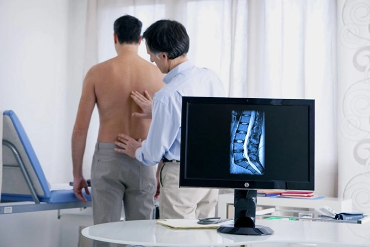
The CT images will be evaluated by one of our neuroradiologists to ensure they are of diagnostic quality. After being monitored for a short time, you will be released with discharge instructions. The entire procedure usually takes about 45-60 minutes.
For your safety and the protection of others, we do not allow anyone other than patients in our exam rooms.
Significant complications related to a myelogram are very uncommon. The primary risks associated with this procedure include:
• Bleeding
• Infection
• Allergic reaction to the contrast material
The most common side effect after a myelogram is a spinal headache, in which leakage of spinal fluid into the surrounding tissues produces a severe headache that typically worsens with standing. Spinal headaches usually resolve on their own, but an additional procedure called an epidural blood patch may be required to stop the leakage of spinal fluid and relieve the symptoms.
Most myelograms are very well tolerated, with minimal discomfort afterwards that is usually easily controlled with non-prescription pain medication. You may apply ice to the needle insertion site to reduce swelling if necessary. Symptoms usually disappear within 48 hours; contact your physician or healthcare provider if they persist for more than two days.
After your study, the images will be evaluated by one of our board-certified radiologists with expertise in neuroradiology. A final report will be sent to your doctor or healthcare provider, who can then discuss the results with you in detail.
Reports are also available on the Patient Portal