
A Dozen Reasons for a Chest CT Scan or MRI
A disease or injury that affects your lungs, heart or chest cavity puts your good health in harm’s way. To help uncover the root of your problem, your provider may prescribe a chest computed tomography (CT) scan or magnetic resonance imaging (MRI) exam.
Conditions Diagnosed with Chest CT Scan or MRI
CT scanners and MRI machines take cross-sectional images of your chest cavity. If necessary, the radiologist can stack these images on top of each other to create three-dimensional images.
While CT scanners rely on an X-ray beam that rotates around you, MRI uses magnets and radio waves.
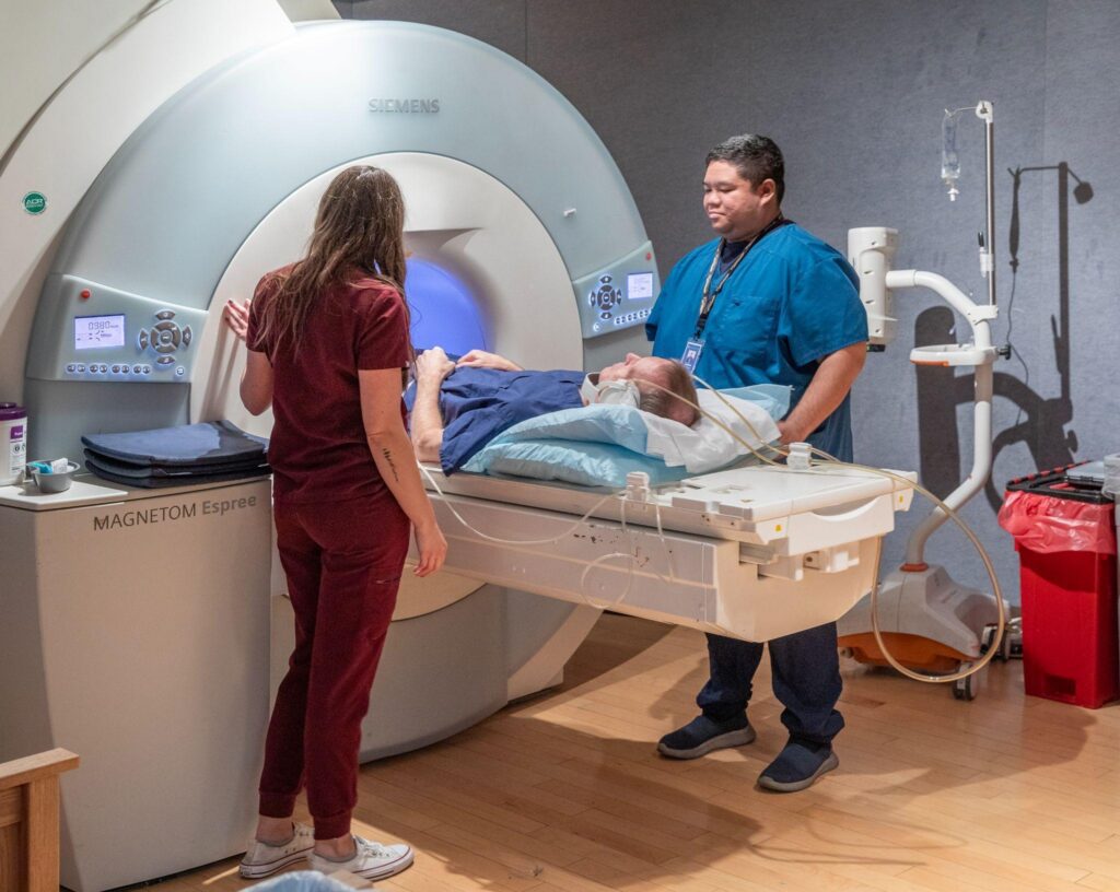
During your exam, your imaging technologist may ask you to hold your breath for short periods of time. This helps capture clear images for accurate diagnosis and treatment planning for many medical conditions.
Here are twelve of them.
#1: Esophageal Cancer and Other Disorders
The esophagus connects the throat to the stomach. If the esophagus develops cancer or other issues, a chest CT scan or MRI can help find the problem.
CT and MRI both help show if cancer spreads beyond the esophagus to the brain, spinal cord or other areas of the body.
#2: Heart Conditions
A cardiac MRI or chest CT scan can identify many heart-related conditions.
Heart conditions that an MRI helps detect include:
- Congenital heart problems (heart defects present at birth)
- Damage caused by a heart attack
- Heart failure
- Problems with your heart valves
- Tumors of the heart
When your provider needs a different view of the heart, a specific type of chest CT scan may help.
- Cardiac computed tomography angiography (CTA) helps identify blood clots by showing how well blood flows through the arteries.
- CT calcium scoring identifies calcium buildup in the heart’s arteries.
#3: Hernias
The diaphragm is a layer of muscle separating your chest from your abdomen. When it weakens, part of your stomach may push through, causing a hiatal hernia.
While X-ray usually diagnoses this issue, a chest CT scan or MRI can provide additional information that’s useful for treatment planning.
#4: Interstitial Lung Disease
Interstitial lung disease (ILD) is a collective term for a group of lung conditions that cause lung inflammation and scarring.
A few types of ILD include:
- Pneumoconiosis
- Pulmonary fibrosis
- Sarcoidosis
- Smoker’s lung
A chest CT scan helps your provider visualize ILD-related damage, track disease progression, and determine if treatment changes are needed moving forward.
#5: Lung Cancer
A low-dose lung CT scan provides lung cancer screening for those at high risk of lung cancer.
The U.S. Preventive Services Task Force recommends this special type of CT every year if you meet the following criteria:
- Age. You are between 50 and 80 years of age.
- Life history. You smoked an average of one pack every day for at least 20 years.
- Recent history. You still smoke or stopped within the past 15 years.
Low-dose lung CT scanning helps detect lung cancer in its earliest stages, before the cancer spreads and causes symptoms.
#6: Mediastinal Tumors
Between your lungs is an area called the mediastinum. Vital nerves and blood vessels are in this area.
- CT with IV contrast can provide detailed information about the tumor.
- MRI can show if the tumor is growing and spreading.
#7: Pleural Disorders
Pleura tissue lines the inside of your chest cavity and the outside of your lungs.
A few pleural conditions include:
- Pleural effusion. Excess fluid builds up in the pleural space.
- Pleurisy. The pleura becomes inflamed.
- Pneumothorax. Air or gas gets trapped in the pleural space.
#8: Pulmonary Embolism
A pulmonary embolism occurs when blood clots develop in a leg, travel to the lungs and become lodged in an artery there.
A pulmonary angiogram helps detect the problem, often using IV contrast to highlight blockages.
#9: Rib and Chest Trauma
Following an automobile accident or other traumatic event affecting your chest, a chest CT scan provides insight into the health of your organs, bones, and soft tissues.
- Blood vessels
- Heart
- Lungs
- Ribs
- Spine
#10: Respiratory Illnesses
COVID-19, flu, pneumonia, and other respiratory illnesses can result in shortness of breath, fever, and fatigue.
When symptoms persist or worsen, a chest CT scan can identify lung damage and help pinpoint the underlying cause.
#11: Thoracic Aortic Aneurysms
An aortic aneurysm occurs when a bulge develops in your aorta. When the bulge occurs inside your chest, it’s a thoracic aortic aneurysm (TAA).
An MRI or chest CT scan can show the size and shape of a TAA.
#12: Thymic Abnormalities
Your thymus is a small gland with a big responsibility. Problems, including benign and cancerous masses, can affect the thymus.
These abnormalities can be detected on a chest X-ray, but a chest CT scan provides more information for diagnosis and treatment.
Chest CT: Valuable in Detecting Disease
A chest CT scan or MRI helps detect problems in the lungs, heart, and surrounding areas. These imaging tests capture detailed pictures that lead to an accurate diagnosis and tailored treatment plan.
CT scanners use X-ray technology, while MRI machines use magnets and radio waves to create cross-sectional images.
Both technologies help diagnose conditions such as heart disease, lung cancer and bulging arteries in the chest area.
Did your provider order a chest CT or MRI for you? Call (520) 733-7226 or request an appointment online with caring experts at your local Radiology Ltd. location.